Brightfield microscopy is attractive because it fits existing lab routines: no dye, no extra channel, and no special acquisition step just to count cells. The tradeoff is that cells can be low-contrast, clustered, or visually different between culture formats. Good software has to handle that variation while still giving scientists a result they can check.
Look for masks, not just numbers
A count without a visible segmentation mask is hard to audit when the assay result matters.
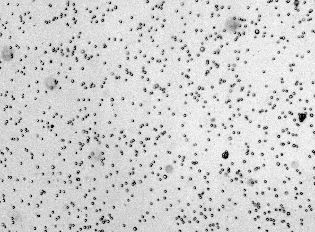
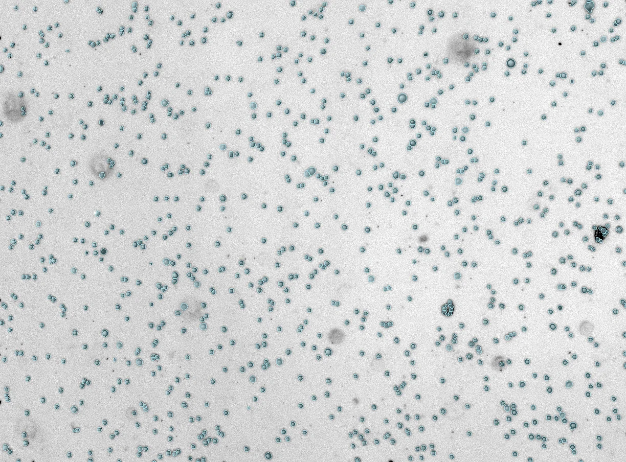 Mask
Raw
Mask
Raw
Features worth checking
The software should fit the experiment, not force every assay into the same generic counting step.
- Segmentation masks that can be reviewed
- Batch upload for plate and time-course images
- CSV exports with image-level measurements
- Support for adherent, suspension, scratch, and spheroid workflows
Warning signs
A fast count can still be the wrong count if the workflow hides quality problems or makes it hard to trace numbers back to images.
- No way to inspect the segmentation
- Manual threshold changes for every batch
- Outputs that need heavy spreadsheet cleanup
Match the tool to the assay
Want to test brightfield images from your lab?
Use CellOpsis in the browser for a quick check, or send a small representative batch if your images need assay-specific tuning.
Try CellOpsis ->