Manual counting is still useful for quick checks, unusual samples, and experiments where a scientist needs to look closely at a few fields of view. It becomes a bottleneck when the same measurement has to be repeated across a plate, a dilution series, or a time-course experiment where consistency matters as much as speed.
Manual counting works when
The dataset is small and the decision depends on a scientist's visual judgement more than a repeatable measurement pipeline.
- One-off checks before passaging
- Small teaching or troubleshooting datasets
- Samples with rare morphology that need close inspection
Automation helps when
Images arrive in batches and you need counts, confluency, masks, and CSV outputs that can be reviewed without redoing the whole analysis by hand.
- 96-well or 384-well plate images
- Time-course experiments with many frames
- Assays where reviewer drift can change conclusions
What changes when you automate?
Scales with every extra image and reviewer.
Scales better across images once the workflow is set.
Can shift with fatigue, threshold choices, and reviewer style.
Uses the same model settings across the batch.
Often captured as a number in a notebook or spreadsheet.
Pairs counts with masks and analysis-ready exports.
The count should come with evidence
Automated counting is strongest when the segmentation can be reviewed before the CSV is trusted.
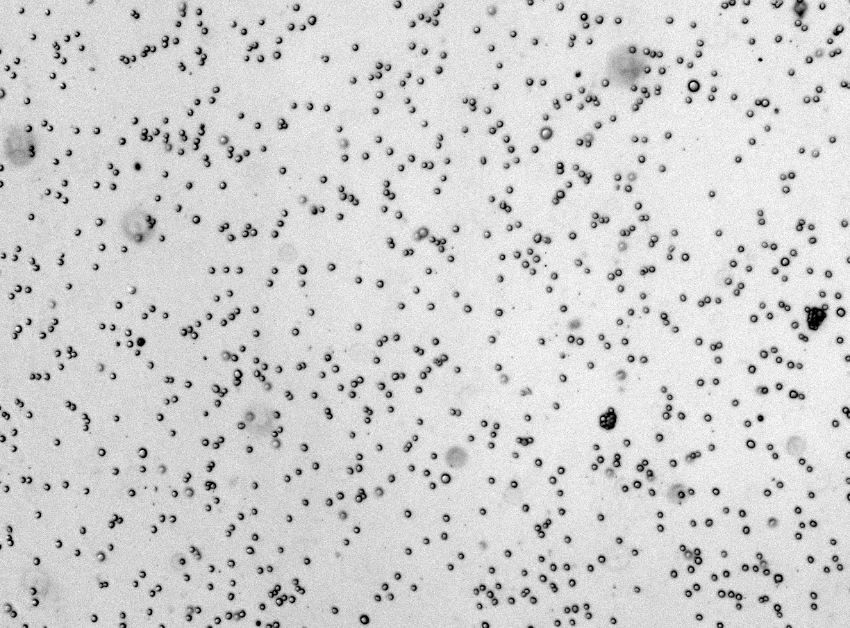
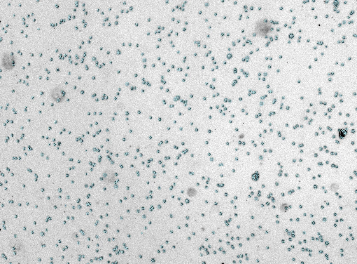 Mask
Raw
Mask
Raw
Have a counting workflow that is eating time?
Try CellOpsis on a representative image, or share a batch if you want to know whether automation will work for your assay.
Try CellOpsis ->